
300+ Appointment
Booking Confirm for
this Week
- Amniotic Membrane Grafting
- Applanation Tonometry
- Concretion Removal
- Corneal Tear Repair
- Corneoscleral Tear Repair
- Cyclocryopexy
- Dressing
- Enucleation/Evisceration
- Foreign Body Removal
- Fundus Fluorescein Angiography
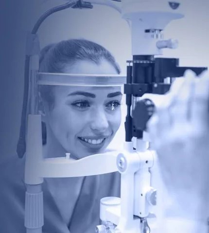
- Glaucoma Work-Up
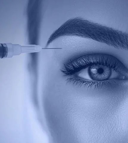
- Intravitreal Injection
- LASIK Work
- Lid Repair
- Meibography
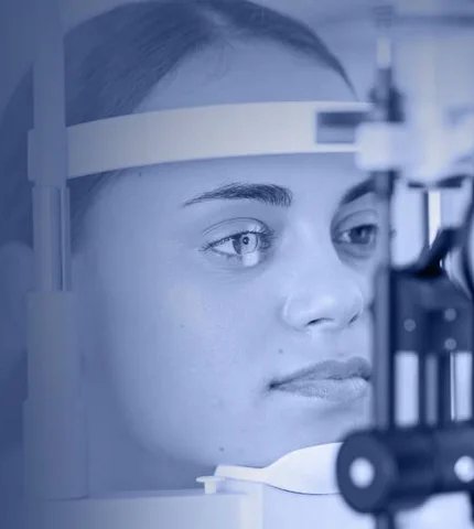
- OCT (Optical Coherence Tomography)
- Ophthalmic Ultrasonography
- Posterior Sub-Tenon Injection
- Pterygium
- Retina Laser
- Retinal Cryopexy
- Retinal Surgeries
- Surgeries
- Topography
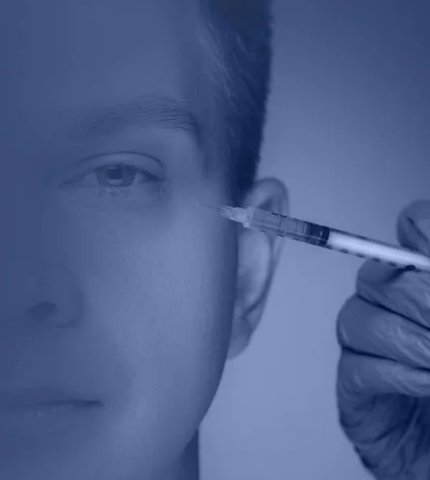
- Supra-Tarsal Injection
- YAG Procedures
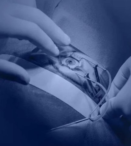
Amniotic Membrane Grafting
Amniotic Membrane Grafting (AMG) is an advanced medical procedure that uses the innermost layer of the placenta, known as the amniotic membrane, to heal and repair damaged tissues in the eye. This natural membrane is rich in healing properties, including growth factors such as Epidermal Growth Factor (EGF) and Transforming Growth Factor-Beta (TGF-β), which stimulate tissue regeneration, reduce inflammation, and minimize scarring.
The amniotic membrane can be used in two primary forms:
- Cryopreserved membranes: Preserved through freezing to retain biological activity, ideal for acute cases requiring high healing potential.
- Dehydrated membranes: Processed through drying for longer shelf-life and ease of use, often chosen for specific planned procedures.
In eye care, AMG is beneficial for treating a variety of conditions, including:
- Corneal ulcers and persistent epithelial defects
- Severe eye injuries such as chemical or thermal burns
- Stevens-Johnson Syndrome – a severe inflammatory condition affecting the eye surface
- Pterygium surgery – to prevent recurrence and improve healing outcomes
- Severe ocular surface disorders, such as conjunctival scarring or limbal stem cell deficiency
By covering the affected area with a protective biological layer, AMG not only reduces pain and inflammation but also promotes epithelialization, improves tissue repair, and restores the eye’s health and function.
The AMG Procedure at Desai Eye Hospital
At Desai Eye Hospital, the AMG procedure is performed by experienced ophthalmologists using proven and advanced techniques:
- Evaluation: A comprehensive eye examination is conducted to assess the condition and determine the suitability of AMG.
- Membrane Preparation: The amniotic membrane, processed under strict medical guidelines to ensure safety and efficacy, is prepared in its cryopreserved or dehydrated form, depending on the condition and treatment plan.
- Application: The membrane is carefully placed over the affected area, tailored to the specific needs of the condition. It may be secured using sutures, fibrin adhesives, or specialized contact lenses.
- Follow-Up Care: Patients receive detailed post-procedure guidance and regular follow-ups to monitor healing and ensure the best outcomes.
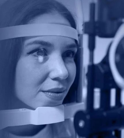
Applanation Tonometry
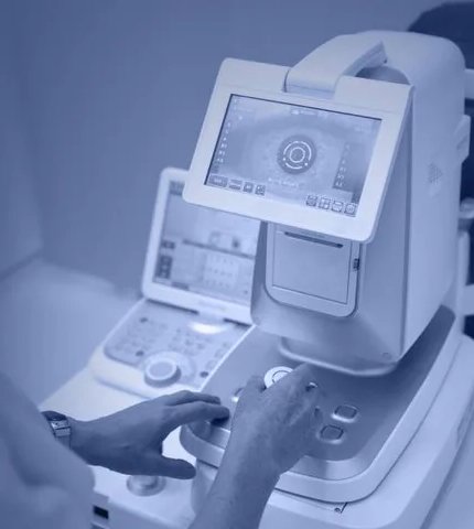
Applanation Tonometry is a widely used diagnostic procedure that measures intraocular pressure (IOP)—the fluid pressure inside the eye. Monitoring IOP is crucial for diagnosing and managing glaucoma, a leading cause of irreversible blindness worldwide. Accurate measurement of eye pressure helps detect abnormalities early and allows for timely intervention to prevent vision loss.
This procedure is based on the principle that the pressure within the eye can be determined by measuring the force required to flatten a specific area of the cornea. It is considered the gold standard for IOP measurement due to its precision and reliability.
Applications of Applanation Tonometry
Applanation Tonometry is essential for:
- Glaucoma Screening: Detecting elevated intraocular pressure, a major risk factor for glaucoma.
- Glaucoma Management: Monitoring IOP regularly to assess the effectiveness of treatments, such as medications, laser therapy, or surgery.
- Ocular Hypertension: Identifying elevated IOP in individuals who may be at risk of developing glaucoma.
- Post-Surgical Monitoring: Ensuring normal eye pressure following ocular surgeries.
The Applanation Tonometry Procedure at Desai Eye Hospital
At Desai Eye Hospital, Applanation Tonometry is performed using state-of-the-art equipment and meticulous care to ensure accurate and comfortable testing:
Concretion Removal
Concretion removal is a specialized ophthalmic procedure used to eliminate tiny calcified deposits or foreign particles embedded in the conjunctiva—the thin, transparent membrane covering the white part of the eye and the inner surface of the eyelids. These deposits, known as concretions, can cause irritation, redness, a foreign body sensation, or discomfort in the eye, affecting the patient’s quality of life.
Applications of Concretion Removal
Concretion removal is performed to:
- Relieve Discomfort: Eliminate irritation and foreign body sensation caused by concretions.
- Prevent Complications: Address calcified deposits before they lead to further inflammation or infection.
- Improve Eye Health: Restore comfort and protect the conjunctiva from damage caused by persistent concretions.
The Concretion Removal Procedure at Desai Eye Hospital
Desai Eye Hospital, concretion removal is a quick and precise outpatient procedure performed with care to ensure patient comfort and safety:
- Preparation: Topical anesthetic drops are applied to numb the eye and ensure a painless experience.
- Inspection: The ophthalmologist carefully examines the conjunctiva using a slit lamp to identify the location and size of the concretions.
- Removal: Using a fine surgical instrument, the concretions are gently extracted without damaging the surrounding tissues.
- Post-Procedure Care: Antibiotic eye drops may be prescribed to prevent infection, and patients are given clear aftercare instructions.
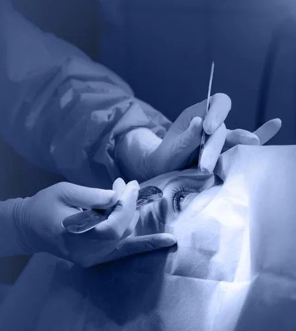
Corneal Tear Repair
Corneal Tear Repair involves the careful suturing or sealing of a damaged cornea caused by trauma, such as lacerations or perforations. The procedure aims to close the wound, restore corneal shape, and maintain its clarity, which is crucial for vision. Depending on the severity of the tear, surgical techniques may include sutures, adhesives, or grafts.
Applications of Corneal Tear Repair
Corneal Tear Repair is essential for:
- Trauma Management: Treating corneal injuries caused by sharp objects, blunt trauma, or foreign bodies.
- Vision Restoration: Ensuring the cornea heals correctly to maintain or restore clear vision.
- Infection Prevention: Closing the tear to reduce the risk of microbial infection, which can cause further complications.
- Ocular Surface Stability: Repairing structural damage to support overall eye health and function.
The Corneal Tear Repair Procedure at Desai Eye Hospital
At Desai Eye Hospital, Corneal Tear Repair is performed by expert ophthalmologists using advanced techniques:
- Assessment: A thorough examination is conducted to evaluate the size, depth, and location of the corneal tear. Imaging tools, such as optical coherence tomography (OCT), may be used for detailed visualization.
- Anesthesia: Local or general anesthesia is administered to ensure patient comfort during the procedure.
- Repair: Depending on the severity of the injury, the cornea is repaired using:
- Sutures: Delicate, high-precision sutures to close the tear.
- Tissue Adhesives: Special adhesives for smaller or superficial tears.
- Grafts: In cases of extensive damage, a corneal graft may be required.
- Post-Operative Care: Patients are prescribed medications to prevent infection, reduce inflammation, and promote healing. Follow-up visits are scheduled to monitor recovery.
Corneoscleral Tear Repair
Corneoscleral tear repair is a critical surgical procedure performed to address injuries that affect both the cornea (the clear, dome-shaped surface of the eye) and the sclera (the white outer layer). Such injuries are typically caused by trauma, and immediate medical attention is crucial to prevent complications such as infection, vision loss, or structural damage to the eye.
This specialized repair procedure aims to restore the integrity of the eye, minimize the risk of further damage, and preserve or improve vision. It requires a high level of precision and expertise to ensure successful outcomes.
Applications of Corneoscleral Tear Repair
Corneoscleral tear repair is essential in the following scenarios:
- Traumatic Eye Injuries: Repairing tears caused by sharp objects, blunt trauma, or accidents.
- Complex Ocular Lacerations: Managing cases where both the cornea and sclera are affected, requiring meticulous reconstruction.
- Preventing Complications: Reducing the risk of infections, structural deformities, and loss of vision caused by untreated eye injuries.
The Corneoscleral Tear Repair Procedure at Desai Eye Hospital
Corneoscleral tear repair is performed by experienced ophthalmic surgeons using advanced techniques and equipment to ensure optimal outcomes:
- Initial Assessment: A thorough examination is conducted to assess the extent of the injury and identify any associated damage within the eye.
- Preparation for Surgery: The patient is prepared for surgery, which is typically performed under local or general anesthesia, depending on the severity of the tear.
- Precise Repair: The surgeon meticulously sutures the cornea and sclera using fine, specialized materials to restore the structural integrity of the eye.
Cyclocryopexy
Cyclocryopexy is a specialized therapeutic procedure designed to manage elevated eye pressure in patients with advanced or treatment-resistant glaucoma. This minimally invasive technique involves applying controlled cold therapy to the ciliary body, a structure in the eye responsible for producing aqueous humor (the fluid within the eye). By reducing fluid production, Cyclocryopexy effectively lowers eye pressure, helping to prevent further damage to the optic nerve and preserve vision.
Applications of Cyclocryopexy
Cyclocryopexy is commonly recommended in cases where other treatments are insufficient or unsuitable, including:
- Advanced Glaucoma: Managing eye pressure in cases of severe glaucoma that have not responded to medications or surgeries.
- Neovascular Glaucoma: Treating elevated eye pressure caused by abnormal blood vessel growth in the eye.
- Refractory Glaucoma: Controlling pressure in patients unresponsive to conventional therapies such as medications, laser treatments, or filtration surgeries.
- Pain Management: Alleviating eye pain associated with uncontrolled glaucoma.
The Cyclocryopexy Procedure at Desai Eye Hospital
At Desai Eye Hospital, Cyclocryopexy is performed with precision and care to ensure effective treatment while prioritizing patient comfort:
- Preparation: The eye is numbed with a local anesthetic to ensure the procedure is painless.
- Cryotherapy Application: A cryoprobe, a specialized instrument that delivers precise freezing temperatures, is gently applied to the outer surface of the eye to target the ciliary body.
- Controlled Treatment: The freezing process reduces the production of aqueous humor, lowering intraocular pressure.
- Post-Procedure Care: Patients are monitored closely and provided with detailed instructions to support recovery and optimize outcomes.
Dressing
Eye dressing involves covering the eye with sterile materials to shield it from external irritants, reduce the risk of infection, and support the healing process. This may include the use of specialized eye pads, bandages, or shields, depending on the patient’s condition and the type of surgery or injury.
Applications of Eye Dressing
Eye dressing is essential in several scenarios:
- Post-Surgical Protection: Safeguarding the eye after procedures such as cataract surgery, corneal transplants, or glaucoma surgery.
- Injury Management: Protecting the eye following trauma or corneal abrasions to prevent further damage.
- Infection Control: Reducing exposure to contaminants that could lead to infections.
- Healing Support: Creating an optimal environment for faster and more effective recovery.
The Eye Dressing Procedure at Desai Eye Hospital
At Desai Eye Hospital, eye dressing is performed with meticulous care and attention to detail:
- Preparation: The eye and surrounding area are cleaned using sterile solutions to minimize the risk of infection.
- Application: A sterile eye pad or bandage is placed over the eye and securely fastened using medical-grade adhesive or a protective eye shield.
- Instructions: Patients are given clear guidance on how to care for the dressing, including when to remove or replace it and signs of complications to watch for.
Why Choose Desai Eye Hospital for Eye Dressing?
- Expert Care: Our experienced team ensures the dressing is applied properly and comfortably for optimal protection and healing.
- High-Quality Materials: We use advanced, sterile materials designed specifically for ophthalmic care.
- Customized Solutions: Eye dressing is tailored to the individual needs of each patient based on their procedure or injury.
Enucleation/Evisceration
Enucleation and Evisceration are surgical procedures used to address severe eye conditions when vision preservation is no longer possible. Enucleation involves removing the entire eye while maintaining the surrounding structures, while Evisceration removes the eye's internal contents, leaving the outer shell and muscles intact. These procedures help manage advanced ocular diseases, relieve pain, and prepare for prosthetic eye placement with a focus on patient comfort.
Applications of Enucleation/Evisceration
These procedures are typically recommended in the following situations:
- Severe Eye Trauma: Addressing irreparable damage caused by traumatic injuries.
- Advanced Ocular Tumors: Removing malignant tumors to prevent further spread and protect overall health.
- Chronic Infections or Inflammation: Treating painful, untreatable infections or conditions like endophthalmitis.
- Painful Blind Eye: Relieving chronic pain in a non-functional eye.
- Cosmetic and Prosthetic Rehabilitation: Preparing the orbit for prosthetic eye placement to restore appearance.
The Enucleation/Evisceration Procedure at Desai Eye Hospital
At Desai Eye Hospital, Enucleation and Evisceration are performed with meticulous care by experienced ophthalmologists:
- Pre-Surgical Assessment: A thorough evaluation is conducted to determine the most suitable procedure based on the patient’s condition.
- Anesthesia: The procedure is performed under local or general anesthesia to ensure patient comfort.
- Surgical Technique:
- In Enucleation, the entire eyeball is carefully removed while preserving the surrounding tissues for future prosthetic placement.
- In Evisceration, the internal contents of the eye are removed while maintaining the scleral shell and muscles.
Foreign Body Removal
Foreign body removal is a critical procedure performed to extract objects that have entered the eye, causing discomfort, irritation, or potential harm. Foreign materials, such as dust, metal, wood, or other small particles, can damage the delicate structures of the eye if not promptly and properly removed. Early intervention is essential to prevent complications, including infections, corneal scratches, or vision impairment.
Applications of Foreign Body Removal
Foreign body removal is necessary for a range of situations, including:
- Superficial Particles: Removal of dust, sand, or similar materials lodged on the cornea or under the eyelid.
- Penetrating Objects: Extraction of sharp or pointed materials such as wood splinters or metal fragments.
- Chemical Debris: Clearing particles from industrial accidents or other exposure to chemical-laden environments.
- Post-Trauma Care: Treatment following accidents involving foreign objects in or around the eye.
The Foreign Body Removal Procedure at Desai Eye Hospital
At Desai Eye Hospital, the foreign body removal procedure is performed with precision and care to ensure patient safety and comfort:
- Evaluation: The affected eye is thoroughly examined using advanced tools such as a slit lamp microscope to locate the foreign object.
- Preparation: Topical anesthetic drops are applied to numb the eye and minimize discomfort during the procedure.
- Removal: Using sterile instruments, the foreign body is carefully extracted from the eye. Depending on its location, a small needle, cotton swab, or specialized tool may be used.
- Cleaning and Aftercare: The eye is rinsed with saline solution to remove any residual particles, and an antibiotic ointment or eye drops may be prescribed to prevent infection and promote healing.
- Follow-Up: Patients are monitored to ensure there is no lingering irritation, infection, or damage to the eye.
Why Choose Desai Eye Hospital for Foreign Body Removal?
- Expert Ophthalmologists: Our team is highly experienced in performing foreign body removal with precision and care.
- Advanced Diagnostic Tools: We use the latest equipment to ensure accurate identification and safe removal of foreign objects.
- Comprehensive Care: From diagnosis to post-procedure follow-up, we provide holistic care to ensure optimal eye health.
Fundus Fluorescein Angiography
Fundus Fluorescein Angiography (FFA) is a diagnostic procedure that uses a special dye injected into the bloodstream to highlight the blood vessels in the retina. When viewed under blue light, it helps detect conditions like diabetic retinopathy, macular degeneration, and retinal vein occlusion. FFA allows ophthalmologists to identify abnormalities such as leakage, blockages, or abnormal blood vessel growth, enabling early intervention and better management of retinal diseases that can lead to vision loss.
Applications of Fundus Fluorescein Angiography
Fundus Fluorescein Angiography is essential for:
- Retinal Disease Diagnosis: Identifying and assessing conditions like diabetic retinopathy, macular degeneration, and retinal vein occlusion.
- Monitoring Diabetic Retinopathy: Evaluating the health of the retina in diabetic patients to detect early signs of damage.
- Age-Related Macular Degeneration (AMD): Detecting abnormal blood vessel growth and retinal damage associated with AMD.
- Retinal Vascular Disorders: Diagnosing conditions related to retinal vessel leakage or blockage, such as retinal vein occlusion.
The Fundus Fluorescein Angiography Procedure at Desai Eye Hospital
At Desai Eye Hospital, Fundus Fluorescein Angiography is performed with the utmost precision and care to ensure optimal results:
- Preparation: A dilating eye drop is applied to widen the pupils, allowing a clear view of the retina.
- Fluorescein Injection: A small amount of fluorescein dye is injected into the arm, and the dye circulates through the bloodstream, highlighting the retinal blood vessels.
- Imaging: Using specialized camera equipment, a series of photographs are taken as the dye moves through the retinal vessels. The dye fluoresces under a blue light, creating detailed images.
- Results and Diagnosis: The images captured during the procedure are carefully analyzed to detect any abnormalities in the retinal blood vessels, which can then guide treatment decisions.
Glaucoma Work-Up
A Glaucoma Work-Up is a comprehensive diagnostic assessment to evaluate the risk of glaucoma and monitor overall eye health. Glaucoma is a leading cause of vision loss, often progressing without noticeable symptoms, making early detection essential. This work-up includes key tests to measure intraocular pressure (IOP), assess optic nerve health, and detect changes in visual fields. Regular monitoring ensures timely intervention, enabling effective management and preservation of vision.
Applications of Glaucoma Work-Up
A comprehensive Glaucoma Work-Up is essential for:
- Glaucoma Screening: Identifying individuals at risk of developing glaucoma based on family history, age, and IOP levels.
- Diagnosis: Detecting early signs of glaucoma before significant damage occurs to the optic nerve.
- Monitoring Disease Progression: Tracking changes in eye pressure, optic nerve condition, and visual fields over time.
- Treatment Planning: Adjusting medications, laser therapies, or surgical interventions based on accurate and thorough evaluations.
The Glaucoma Work-Up Procedure at Desai Eye Hospital
At Desai Eye Hospital, our glaucoma work-up is performed with the utmost care and precision using the latest diagnostic tools:
- Initial Assessment: The ophthalmologist conducts a thorough eye examination, discussing your medical history and risk factors for glaucoma.
- Intraocular Pressure Measurement: We measure the pressure inside the eye using advanced tonometry techniques, such as Applanation Tonometry, to detect any elevated IOP levels.
- Optic Nerve Imaging: Using high-resolution imaging technologies, we assess the optic nerve to check for signs of damage that may indicate glaucoma.
- Visual Field Test: A visual field test helps identify any peripheral vision loss, which is common in glaucoma patients.
- Pachymetry: This test measures the thickness of the cornea, as thinner corneas can increase the risk of developing glaucoma.
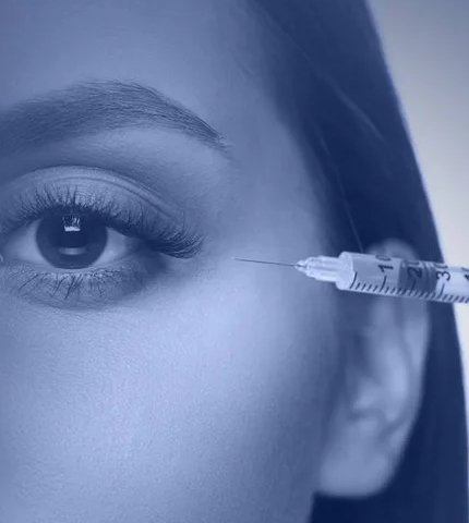
Intravitreal Injection
Intravitreal injections are an effective treatment for serious retinal conditions like macular degeneration, diabetic retinopathy, and retinal vein occlusion. The procedure involves delivering medication directly into the vitreous (the gel inside the eye) to target and treat these conditions, helping to preserve vision and prevent further damage.
This safe, outpatient procedure provides concentrated medication exactly where it's needed, offering fast and effective results with expert care.
Applications of Intravitreal Injection
Intravitreal injections are essential for treating the following conditions:
- Age-Related Macular Degeneration (AMD): A leading cause of vision loss in older adults, requiring prompt treatment to slow progression and preserve central vision.
- Diabetic Retinopathy: A complication of diabetes that can lead to blindness, managed with injections to reduce swelling and prevent further damage.
- Retinal Vein Occlusion (RVO): A blockage of the retinal veins that can lead to vision loss, treated with injections to reduce inflammation and improve circulation.
- Macular Edema: Swelling in the retina, often associated with other eye conditions, treated with medication delivered directly to the site.
The Intravitreal Injection Procedure at Desai Eye Hospital
At Desai Eye Hospital, we perform intravitreal injections with the utmost care and precision using advanced techniques:
- Preparation:The area around the eye is cleaned thoroughly, and a local anesthetic is applied to ensure comfort throughout the procedure.
- Injection:A tiny needle is used to inject the medication into the vitreous, targeting the affected area of the retina. The procedure is quick and typically takes only a few minutes.
- Post-Procedure Care: After the injection, patients are monitored briefly to ensure there are no immediate complications. Instructions for aftercare, including using prescribed eye drops, will be provided.
LASIK Work
LASIK (Laser-Assisted in Situ Keratomileusis) is a highly effective procedure that corrects refractive vision problems, including nearsightedness, farsightedness, and astigmatism. By reshaping the cornea with a precise laser, LASIK improves how light is focused on the retina, providing clearer vision without the need for glasses or contact lenses. Known for its high success rate, LASIK offers a safe and reliable solution for permanent vision correction.
Applications of LASIK Surgery
- Nearsightedness (Myopia): Correcting vision in individuals who have difficulty seeing distant objects.
- Farsightedness (Hyperopia): Improving vision for those who struggle with seeing nearby objects.
- Astigmatism: Addressing blurred or distorted vision caused by an irregularly shaped cornea.
The LASIK Procedure at Desai Eye Hospital
At Desai Eye Hospital, LASIK is performed using advanced technology and precision for optimal results:
- Preparation: The eye is numbed with topical anesthetic drops to ensure a pain-free procedure.
- Flap Creation: A thin flap is created in the cornea using a laser or microkeratome, allowing access to the inner corneal tissue.
- Laser Treatment: The excimer laser is used to reshape the cornea, correcting the refractive error. This process is quick and minimally invasive.
- Flap Repositioning: After the cornea is reshaped, the flap is carefully repositioned, where it naturally adheres without the need for stitches.
- Recovery: The procedure is fast, typically lasting only 15-20 minutes per eye, with minimal discomfort during recovery.
Why Choose Desai Eye Hospital for LASIK?
- Precision and Expertise: Our skilled ophthalmologists utilize the latest LASIK technology for highly accurate and customized treatment.
- Advanced Equipment: We use cutting-edge lasers and diagnostic tools to ensure optimal results and patient safety.
- Personalized Care: We offer comprehensive pre-surgical assessments to ensure LASIK is the right choice for your eyes and vision needs.
Lid Repair
Lid Repair is a specialized surgical procedure to treat the eyelids' injuries, deformities, or abnormalities. The eyelids are essential for protecting the eyes, maintaining tear distribution, and supporting vision. Conditions such as trauma, congenital defects, or eyelid drooping can impact both function and appearance.
Lid Repair reconstructs the eyelid, addressing lacerations, drooping, or other issues that affect eyelid movement and closure. This precise procedure restores the eyelids' structure and function, ensuring minimal scarring and optimal eye health.
Applications of LID Repair
LID Repair is essential for:
- Traumatic Injuries: Repairing eyelid lacerations or damage from accidents.
- Congenital Abnormalities: Correcting birth defects like ptosis (drooping eyelids) or ectropion (outward-turning eyelids).
- Tumor Removal: Reconstructing the eyelid after the excision of tumors or growths.
- Cosmetic Concerns: Addressing cosmetic issues such as eyelid drooping that may affect vision or appearance.
- Functional Repairs: Restoring the ability of the eyelids to properly close, protecting the eye from dryness or exposure.
The LID Repair Procedure at Desai Eye Hospital
At Desai Eye Hospital, LID Repair is performed using advanced surgical techniques to ensure optimal results:
- Preparation: The eye area is numbed using local anesthesia to ensure a painless procedure.
- Surgical Repair: The surgeon carefully reconstructs the eyelid, whether through stitching lacerations, repositioning tissues, or addressing other abnormalities.
- Post-Procedure Care: After the surgery, patients are provided with instructions for proper care and healing to ensure minimal scarring and optimal eyelid function.
- Recovery: Most patients recover with minimal downtime, with follow-up visits to monitor healing and ensure the best cosmetic and functional outcome.
Meibography
Meibography is a non-invasive diagnostic procedure that examines the meibomian glands in the eyelids, which are crucial for producing the oily layer of the tear film. These glands help prevent tear evaporation and maintain eye comfort. When damaged or dysfunctional, they can lead to dry eye disease, causing discomfort and potential vision issues. Meibography provides detailed imaging to detect gland dysfunction early, enabling effective treatment for managing dry eye symptoms.
Applications of Meibography
Meibography is essential for:
- Dry Eye Diagnosis: Identifying meibomian gland dysfunction (MGD), a leading cause of dry eye disease.
- Management of Chronic Dry Eye: Monitoring the health of the meibomian glands to guide treatment plans, including the use of lubricants, medications, or lifestyle changes.
- Pre-Surgical Assessment: Evaluating gland health before ocular surgeries, such as LASIK, to prevent complications related to dry eyes.
- Monitoring Treatment Progress: Tracking improvements or further damage to the meibomian glands during ongoing treatment for dry eyes.
The Meibography Procedure at Desai Eye Hospital
At Desai Eye Hospital, we use advanced imaging technology for Meibography to ensure precise and detailed assessment of your meibomian glands:
- Preparation: The procedure is non-invasive, with no need for anesthetic drops. The patient may be asked to blink a few times to stimulate tear production.
- Imaging: A specialized device is used to capture high-resolution images of the meibomian glands. The patient will be asked to look at a light source while the device takes pictures of the lower and upper eyelids.
- Analysis: The images are analyzed by our skilled ophthalmologists to assess the condition of the meibomian glands, including their structure and function.
- Results and Diagnosis: Based on the findings, a treatment plan is developed to manage dry eye disease and restore gland function where possible.
Why Choose Desai Eye Hospital for Meibography?
- Advanced Technology: We use the latest imaging equipment to provide the most accurate and detailed assessment of your meibomian glands.
- Expert Ophthalmologists: Our team of experienced ophthalmologists specializes in diagnosing and managing dry eye disease with personalized care.
OCT (Optical Coherence Tomography)
Optical Coherence Tomography (OCT) is a non-invasive imaging technique that provides high-resolution images of the retina and optic nerve. It allows for detailed analysis of the eye’s internal structures, aiding in the early detection of conditions like macular degeneration, diabetic retinopathy, and glaucoma. OCT uses light waves to capture cross-sectional views of the retina, helping ophthalmologists monitor and diagnose various ocular conditions with precision, ultimately preventing vision loss.
Applications of OCT
OCT is essential for:
- Macular Disease Diagnosis and Monitoring: Detecting and tracking conditions like age-related macular degeneration (AMD) and diabetic macular edema (DME).
- Glaucoma Management: Evaluating the optic nerve and retinal nerve fiber layer thickness to monitor glaucoma progression.
- Retinal Health Evaluation: Assessing the retina for diseases like diabetic retinopathy and retinal vein occlusion.
- Pre- and Post-Surgical Assessment: Monitoring retinal changes before and after surgeries such as cataracts or retinal procedures.
The OCT Procedure at Desai Eye Hospital
At Desai Eye Hospital, the OCT procedure is performed with precision and care using the latest equipment:
- Preparation: The patient is seated comfortably, and the eye to be examined is positioned in front of the OCT scanner.
- Imaging: The technician will instruct the patient to focus on a specific point while the OCT device captures detailed images of the retina and optic nerve.
- Analysis: The images are analyzed by our ophthalmologists to assess the health of the retina, optic nerve, and other internal structures.
- Results and Diagnosis: The results are available immediately, and the ophthalmologist will discuss the findings and provide recommendations for any necessary treatment or follow-up.
Why Choose Desai Eye Hospital for OCT?
- Advanced Technology: We use the latest OCT equipment to provide the most accurate and detailed imaging available.
- Experienced Ophthalmologists: Our skilled professionals have extensive experience in interpreting OCT images and diagnosing various eye conditions.
Ophthalmic Ultrasonography
Ophthalmic Ultrasonography is an advanced diagnostic technique that uses high-frequency sound waves to visualize the internal structures of the eye and surrounding tissues. This precise imaging method is essential for detecting hidden abnormalities, planning treatments, and monitoring the progression of various eye conditions, ensuring accurate diagnosis and effective management.
Applications of Ophthalmic Ultrasonography
- Detecting Ocular Pathologies: Identifying conditions such as tumors, retinal detachments, vitreous hemorrhages, or foreign bodies within the eye.
- Cataract and Glaucoma Evaluation: Assessing the anterior and posterior segments of the eye, especially in cases where opacities obscure detailed examination.
- Pre-Surgical Planning: Providing valuable insights for procedures such as cataract or retinal surgery.
- Orbital Disorders: Evaluating orbital tumors, abscesses, or other abnormalities in the eye socket.
The Ophthalmic Ultrasonography Procedure at Desai Eye Hospital
At Desai Eye Hospital, ophthalmic ultrasonography is performed with precision and care, ensuring accurate results:
- Preparation: The patient is comfortably positioned, and a numbing eye drop may be applied if necessary.
- Ultrasound Imaging: Using a handheld probe, the ophthalmologist gently scans the eye, sending sound waves that create real-time images of the internal structures.
- Analysis and Diagnosis: The images are evaluated to identify abnormalities or confirm diagnoses, aiding in the creation of a tailored treatment plan.
Why Choose Desai Eye Hospital for Ophthalmic Ultrasonography?
- Advanced Technology: We use high-resolution ultrasound devices to provide clear, detailed images for accurate diagnosis.
- Experienced Ophthalmologists: Our skilled specialists have extensive expertise in interpreting ultrasound findings and formulating effective treatment strategies.
Posterior Sub-Tenon Injection
Posterior Sub-Tenon Injection is a specialized treatment procedure used to deliver medication directly to the back of the eye. This technique is particularly effective in managing inflammatory and retinal conditions by providing targeted therapy with minimal systemic side effects. It is widely recognized for its precision and ability to achieve high concentrations of medication in the desired area, improving treatment outcomes.
What is Posterior Sub-Tenon Injection?
The procedure involves injecting medication into the space beneath the Tenon’s capsule, a thin layer of tissue that envelops the eyeball. This approach ensures that the medication reaches the posterior segment of the eye, where conditions such as macular edema, uveitis, and diabetic retinopathy often occur.
Applications of Posterior Sub-Tenon Injection
Posterior Sub-Tenon Injection is used to manage a variety of eye conditions, including:
- Uveitis: Reduces inflammation in the back of the eye to prevent vision loss.
- Macular Edema: Treats swelling in the retina associated with diabetes, vein occlusion, or other causes.
- Diabetic Retinopathy: Delivers anti-inflammatory or anti-VEGF medication to manage complications.
- Post-Surgical Inflammation: Controls inflammation after procedures like cataract or retinal surgery.
The Posterior Sub-Tenon Injection Procedure at Desai Eye Hospital
At Desai Eye Hospital, Posterior Sub-Tenon Injections are performed with precision and care by our expert ophthalmologists:
- Preparation: The eye is numbed with anesthetic drops to ensure a painless experience.
- Sterilization: The surrounding area is cleaned thoroughly to maintain a sterile environment.
- Injection: Using a fine cannula, the medication is carefully injected into the space beneath the Tenon’s capsule, targeting the affected area.
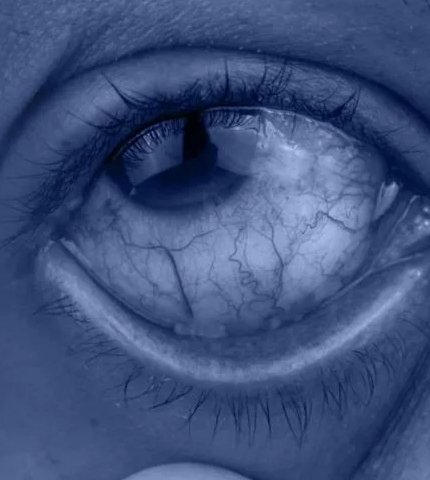
Pterygium
Pterygium is a non-cancerous growth of pink, fleshy tissue on the conjunctiva, the clear membrane covering the white of the eye. While usually harmless, it can cause redness, discomfort, and even vision issues if it extends onto the cornea.
Causes of Pterygium
Pterygium is commonly linked to prolonged exposure to UV light, dry or windy environments, and irritants like dust. It often affects individuals who spend significant time outdoors without proper eye protection.
Prompt diagnosis and treatment are essential to prevent complications and maintain eye health.
Applications of Pterygium Surgery
Pterygium removal is performed to address various symptoms and complications, including:
- Vision Impairment: Removing the tissue when it encroaches on the cornea and affects vision.
- Cosmetic Concerns: Improving the eye’s appearance by removing visible growths.
- Persistent Discomfort: Alleviating irritation, redness, and dryness caused by the growth.
- Preventing Recurrence: Using advanced surgical techniques to reduce the likelihood of regrowth.
The Pterygium Surgery Procedure at Desai Eye Hospital
At Desai Eye Hospital, pterygium surgery is performed using meticulous techniques and advanced care to ensure successful outcomes:
- Evaluation and Planning: A comprehensive eye examination is conducted to assess the size, location, and severity of the pterygium.
- Surgical Removal: The pterygium is carefully excised under local anesthesia to ensure precision and patient comfort.
- Conjunctival Autograft: A graft of healthy conjunctival tissue is taken from another part of the eye and transplanted to the affected area. This technique minimizes the risk of recurrence and promotes healing.
Retina Laser
Retina laser treatment is a highly effective and precise procedure used to manage various retinal conditions that can threaten vision. By delivering controlled laser energy to targeted areas of the retina, this procedure helps stabilize or improve vision and prevent further complications. Retina laser therapy is a cornerstone of modern ophthalmology, offering safe and reliable solutions for managing retinal disorders.
Applications of Retina Laser Treatment
Retina laser treatment is essential for addressing a range of retinal conditions, including:
- Diabetic Retinopathy: Reduces abnormal blood vessels and prevents further damage to the retina caused by diabetes.
- Retinal Tears and Detachments: Seals retinal tears or holes to prevent progression to a retinal detachment.
- Age-Related Macular Degeneration (AMD): Slows the progression of certain forms of AMD by targeting and sealing abnormal blood vessels.
- Vascular Occlusions: Treats complications arising from blocked retinal veins or arteries.
- Macular Edema: Reduces fluid accumulation in the macula to restore central vision.
The Retina Laser Procedure at Desai Eye Hospital
At Desai Eye Hospital, retina laser treatment is performed with precision and care, ensuring patient safety and comfort at every step:
- Preparation: The eye is numbed with anesthetic drops, and the pupil may be dilated for better access to the retina.
- Laser Delivery: Using advanced laser systems, targeted laser energy is applied to the affected area of the retina under the guidance of an experienced ophthalmologist.
- Monitoring: The procedure is carefully monitored to ensure accuracy and effectiveness, with minimal discomfort to the patient.
Retinal Cryopexy
Retinal Cryopexy is a specialized procedure used to treat retinal tears and holes, which, if left untreated, can lead to retinal detachment and vision loss. This procedure utilizes extreme cold to create a scar around the damaged area of the retina, effectively sealing it and preventing further damage. It is a safe, effective, and widely used treatment for stabilizing the retina and preserving vision.
Applications of Retinal Cryopexy
Retinal Cryopexy is essential for:
- Retinal Tear Treatment: Repairing tears in the retina to prevent detachment.
- Retinal Detachment Prevention: Securing areas of weakness to stop further progression.
- Post-Surgical Support: Strengthening retinal repair following other procedures like scleral buckling or vitrectomy.
- Treatment of Lattice Degeneration: Addressing areas of thinning or atrophy in the retina to minimize risks of tears.
The Retinal Cryopexy Procedure at Desai Eye Hospital
At Desai Eye Hospital, Retinal Cryopexy is performed with precision and care by our expert ophthalmologists:
- Preparation: The eye is numbed with topical or local anesthesia to ensure patient comfort.
- Cryotherapy Application: A specialized cryoprobe is used to deliver controlled freezing to the affected area. The extreme cold stimulates the formation of scar tissue, which seals the tear or hole.
- Post-Treatment Care: Patients receive guidance on post-procedure care and follow-up visits to monitor healing and ensure optimal outcomes.
Why Choose Desai Eye Hospital for Retinal Cryopexy?
- Expertise and Experience: Our ophthalmologists are highly skilled in retinal procedures, ensuring precise and effective treatment.
- Advanced Technology: We use the latest cryotherapy equipment to deliver safe and reliable results.
- Comprehensive Care: Retinal Cryopexy is part of our integrated approach to diagnosing and managing retinal conditions.
Retinal Surgeries
Retinal surgeries are advanced procedures used to treat conditions like retinal detachments, macular holes, diabetic retinopathy, and age-related macular degeneration (AMD). The retina plays a vital role in vision by capturing light and transmitting signals to the brain. Timely surgical intervention helps preserve sight and prevent permanent vision loss, using modern techniques and technology for precise and effective outcomes.
Applications of Retinal Surgeries
Retinal surgeries are crucial for managing various conditions, including:
- Retinal Detachment Repair: Reattaching the retina to prevent vision loss.
- Macular Hole Surgery: Sealing holes in the macula to restore central vision.
- Diabetic Retinopathy Treatment: Repairing damage caused by diabetes-related retinal changes.
- Epiretinal Membrane Removal: Treating wrinkling or scarring on the retina.
- Age-Related Macular Degeneration (AMD): Managing complications like bleeding or fluid accumulation.
The Retinal Surgery Procedure at Desai Eye Hospital
At Desai Eye Hospital, retinal surgeries are performed by skilled ophthalmologists using advanced techniques and cutting-edge technology to ensure the best possible outcomes:
- Comprehensive Evaluation: A detailed retinal examination is conducted using diagnostic tools such as optical coherence tomography (OCT) and fundus imaging to assess the condition.
- Surgical Planning: Based on the diagnosis, a personalized surgical plan is developed, which may include vitrectomy, laser photocoagulation, or scleral buckling.
- Precise Execution: Retinal surgeries are performed in a sterile environment using high-precision instruments and imaging guidance to target the affected areas with accuracy.
- Post-Surgery Care: After surgery, patients receive detailed care instructions and regular follow-ups to monitor healing and ensure long-term success.
Surgeries
At Desai Eye Hospital, we offer a range of advanced eye surgeries designed to address various eye conditions and improve your vision. Whether you are dealing with cataracts, glaucoma, or other complex eye issues, our experienced ophthalmologists utilize the latest techniques and technologies to provide effective solutions.
Types of Eye Surgeries at Desai Eye Hospital
We provide the following advanced surgical treatments:
- Cataract Surgery: Removal of the clouded lens and replacement with a clear, artificial lens to restore vision.
- Glaucoma Surgery: A variety of surgical options to reduce eye pressure and manage glaucoma, preventing further damage to the optic nerve.
- Refractive Surgery (LASIK): Corrects nearsightedness, farsightedness, and astigmatism to reduce or eliminate the need for glasses or contact lenses.
- Retinal Surgery: Treats retinal conditions such as retinal detachment, macular degeneration, and diabetic retinopathy.
- Corneal Transplant: Replaces a damaged or diseased cornea with a healthy donor cornea to restore vision.
- Pterygium Surgery: Removes growths from the eye surface to improve vision and comfort.
The Surgical Procedure at Desai Eye Hospital
Each surgery is tailored to the specific needs of the patient and is conducted with the utmost care and precision:
- Pre-Surgery Consultation: Our specialists thoroughly evaluate your condition to determine the best surgical approach.
- Anesthesia: Depending on the type of surgery, local or general anesthesia is administered to ensure comfort throughout the procedure.
- The Surgery: The procedure is performed with advanced surgical instruments, guided by years of expertise, to ensure the best possible outcome.
- Post-Surgery Care: Our team provides detailed aftercare instructions and schedules follow-up visits to monitor healing and ensure the success of the surgery.
Why Choose Desai Eye Hospital for Eye Surgeries?
- Expert Surgeons: Our highly skilled ophthalmologists have extensive experience in performing a wide range of eye surgeries, ensuring the highest level of care and successful outcomes.
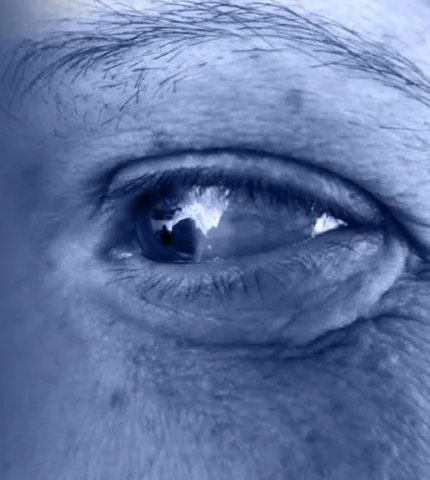
Topography
Topography is an advanced diagnostic procedure that maps the surface of the cornea, providing detailed, three-dimensional images of its curvature. This is essential for diagnosing conditions like corneal irregularities, astigmatism, and vision problems related to the eye's shape. It is especially important for patients considering refractive surgeries such as LASIK, as it helps tailor the procedure to the unique shape of their cornea. The procedure works by analyzing light reflections from the corneal surface, creating an accurate map crucial for treatment planning.
Applications of Corneal Topography
Corneal topography is essential for:
- Diagnosing Corneal Disorders: Identifying conditions like keratoconus, corneal dystrophies, and astigmatism.
- Pre-Surgical Assessment: Evaluating the cornea’s shape before refractive surgeries like LASIK to ensure safe and effective outcomes.
- Monitoring Disease Progression: Tracking changes in corneal shape in patients with keratoconus or after eye surgery.
- Custom Contact Lens Fitting: Assisting in the fitting of custom contact lenses for patients with irregular corneas or vision issues.
The Corneal Topography Procedure at Desai Eye Hospital
At Desai Eye Hospital, corneal topography is performed with precision using the latest technology to ensure accurate and detailed measurements:
- Preparation: The eye is cleaned, and the patient is comfortably seated in front of the topographer machine.
- Mapping: The patient is asked to look at a target light while the topographer scans the cornea’s surface. This process is quick and non-invasive.
- Results and Diagnosis: The machine generates a detailed map of the cornea, which is analyzed by our expert ophthalmologists to provide insights into any irregularities or conditions.
Supra-Tarsal Injection
A Supra-Tarsal Injection is an advanced procedure used to deliver medication directly into the area above the tarsal plate of the eyelid, targeting specific eye conditions. This technique is commonly used to treat various ocular inflammatory conditions, including allergic conjunctivitis, uveitis, and other inflammatory diseases of the eye. Administering the medication directly to the site of inflammation, provides fast and effective relief, reducing symptoms and improving eye health.
Applications of Supra-Tarsal Injection
Supra-tarsal injection is commonly used for:
- Allergic Conjunctivitis: To reduce inflammation and allergic reactions in the eye.
- Uveitis: To manage inflammation of the uvea and prevent complications that could lead to vision loss.
- Ocular Inflammatory Conditions: Treating a variety of inflammatory diseases affecting the anterior or posterior segments of the eye.
- Post-Surgical Inflammation: Used to control inflammation after eye surgeries such as cataract surgery or corneal transplants.
The Supra-Tarsal Injection Procedure at Desai Eye Hospital
At Desai Eye Hospital, the Supra-Tarsal Injection procedure is performed with precision and patient comfort in mind:
- Preparation: The area around the eye is cleaned, and a topical anesthetic is applied to minimize discomfort during the procedure.
- Injection: The ophthalmologist carefully injects the medication into the supra-tarsal space, just above the tarsal plate, using a fine needle. The injection is quick, with minimal discomfort.
- Post-Procedure Care: After the injection, patients may experience mild discomfort or pressure in the eye, which typically resolves quickly. Follow-up visits are scheduled to monitor progress and ensure the medication is working effectively.
Why Choose Desai Eye Hospital for Supra-Tarsal Injection?
- Expert Care: Our skilled ophthalmologists have extensive experience in performing Supra-Tarsal Injections, ensuring the procedure is done safely and effectively.
- Targeted Treatment: We provide personalized care, selecting the most appropriate medications to treat your specific eye condition.
- Comprehensive Eye Health: As part of our full spectrum of ophthalmic services, the Supra-Tarsal Injection contributes to our commitment to improving your eye health and treating various ocular conditions.
YAG Procedures
YAG (Yttrium-Aluminum-Garnet) laser procedures are advanced, non-invasive treatments used to manage eye conditions like cataracts and glaucoma. These procedures effectively improve vision and alleviate symptoms by precisely targeting and treating specific areas of the eye with minimal discomfort. Common applications include capsulotomy for post-cataract surgery patients and trabeculoplasty for glaucoma management. YAG laser treatments offer rapid recovery, making them a reliable option for enhancing eye health and quality of life.
Applications of YAG Procedures
YAG laser procedures are essential for:
- Capsulotomy: Treating posterior capsule opacification (PCO) that may develop after cataract surgery.
- Glaucoma Treatment: Reducing eye pressure by improving the drainage of fluid in the eye, especially in patients with open-angle glaucoma.
- Peripheral Iridotomy: Creating a small hole in the iris to treat angle-closure glaucoma, allowing for proper fluid flow within the eye.
- Vitreolysis: Addressing floaters in the vitreous by breaking them up with the laser, improving vision in some patients.
The YAG Procedure at Desai Eye Hospital
At Desai Eye Hospital, YAG procedures are performed with precision and care to ensure the best results and patient comfort:
- Preparation: The eye is numbed with topical anesthetic drops, ensuring comfort throughout the procedure.
- Laser Application: Using a focused YAG laser, the ophthalmologist targets the specific area of the eye requiring treatment, whether it’s the posterior capsule, drainage angle, or iris.
- Minimal Discomfort: The procedure is minimally invasive, and most patients experience little to no discomfort during the process.
- Results and Follow-Up: The results are immediate, with many patients experiencing an improvement in symptoms or vision. Follow-up care is provided to monitor progress and ensure optimal outcomes.
Our Specialist Doctors
Offering Advanced Eye Care Solutions:
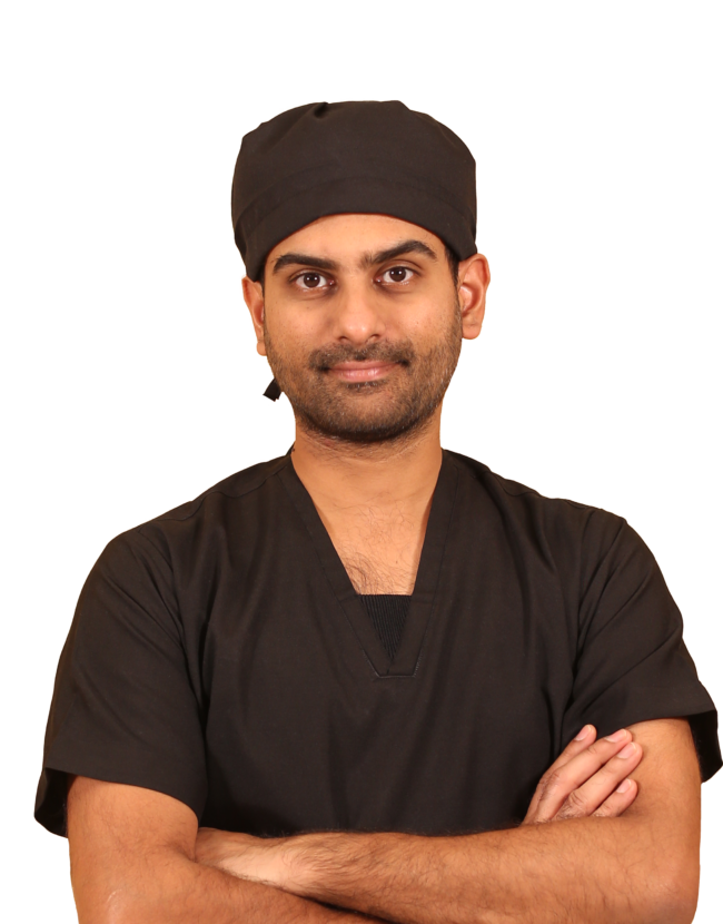
Dr Ankit Desai
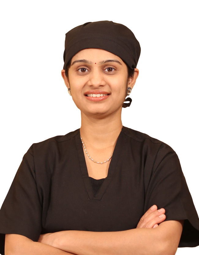
Dr Avani Desai

Dr Sushma Desai

Dr Priya Desai

Dr Sujit Desai


The surgery itself was quick and smooth, and my vision improved significantly within a short time. The clinic maintained excellent hygiene and used advanced equipment, which gave me confidence in the treatment. Post-operative care and guidance were also very helpful.
I would highly recommend Dr. Priya Desai and Desai Eye Institute to anyone considering LASIK or other eye treatments.

My management has been entirely through prescribed eye drops, without surgical intervention.
Both doctors are attentive, patient, and thorough in their explanations.
They took time to help me understand how Fuch’s Dystrophy progresses and why eye drops are the right approach for my current stage.
I appreciated that they didn’t rush me toward surgery, but instead focused on stabilizing my condition with conservative treatment.
Regular check ups are scheduled to monitor corneal clarity and vision changes, which reassures me that my progress is being tracked carefully.
The prescribed drops help reduce morning blurriness and corneal swelling, making daily vision clearer.
I noticed gradual improvement in comfort and reduced glare sensitivity, which has made daily tasks easier.
Doctors are approachable and explain every detail in simple language.
Staff support is smooth, from appointment booking to pharmacy guidance.
The hospital environment is clean and organized, which adds confidence.
My treatment journey for Fuch's Dystrophy under Dr. Sujit Desai and Dr. Priya Desai has been reassuring and effective. Their conservative approach shows genuine care for patient well being, avoiding unnecessary procedures while ensuring close monitoring. I feel confident continuing my care at Desai Eye Institute, knowing that if my condition progresses, advanced options are available—but for now, the drops are working well.
Desai Eye Institute is a reliable choice for comprehensive eye care. Whether it’s routine eye testing or advanced surgical treatment, the hospital delivers professional service with patient satisfaction as a priority. Highly recommended for individuals and families seeking quality eye treatment.

A special and heartfelt thank you to Dr. Priya Desai for her expertise, calm approach, and excellent care.

Prior to the procedure, Dr. Desai conducted a thorough eye examination and performed all the required diagnostic tests. She ensured that LASIK was a safe and appropriate option for me before moving forward. She explained every step of the procedure clearly and patiently, addressing all my concerns and setting realistic expectations.
What I appreciated most was her calm and reassuring approach. She took the time to make me feel comfortable, repeatedly reassuring me and creating a stress-free environment. Her confidence and clarity made a significant difference in easing my nerves.
The staff at Desai Eye Institute were equally impressive—efficient, courteous, and highly supportive at every stage, from testing to surgery and follow-up care. The clinic clearly maintains high standards of organization and patient-centered care.
I am extremely satisfied with my results and overall experience. I highly recommend Desai Eye Institute and Dr. Priya Desai to anyone seeking advanced eye care or LASIK treatment.