
Modern eye care relies heavily on advanced imaging techniques to detect diseases early. One such important diagnostic tool is fundus photography. Many patients ask, what is fundus photography, and how this test helps in identifying serious eye conditions.
In this article, we will explain what is fundus photography, its purpose, how it works, and why it plays a crucial role in preventing vision loss.
What Is Fundus in Eye?
Before understanding the test, it is important to clarify what is fundus in eye.
The fundus is the interior surface at the back of the eye. It includes:
- Retina
- Optic disc
- Macula
- Retinal blood vessels
This area is essential for vision because it processes light signals and sends them to the brain. Any damage to the fundus can directly affect eyesight.
What Is Fundus in Ophthalmology?
In medical terms, what is fundus in ophthalmology refers to the anatomical structures visible during a detailed retinal examination. Ophthalmologists examine the fundus to identify signs of:
- Diabetic retinopathy
- Hypertensive retinopathy
- Glaucoma
- Age related macular degeneration
- Retinal detachment
Since many retinal diseases develop without early symptoms, fundus evaluation is critical for early detection.
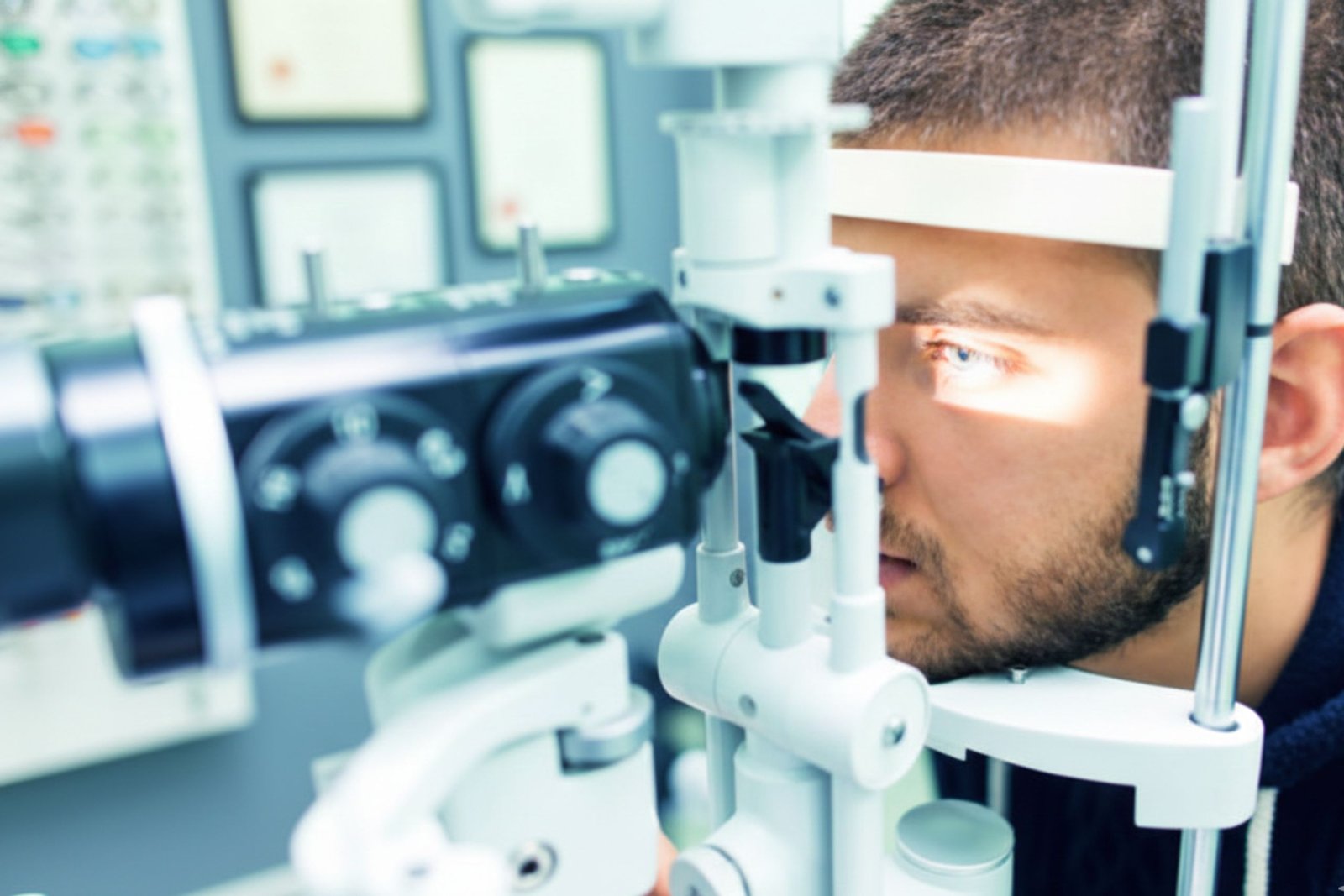
What Is Fundus Photography?
Now let us address the main question, what is fundus photography?
Fundus photography is a non invasive imaging test that captures high resolution photographs of the retina and other structures at the back of the eye. A special fundus camera is used to take detailed color images.
The test helps doctors:
- Document retinal changes
- Monitor disease progression
- Compare images over time
- Detect early abnormalities
According to the World Health Organization, diabetic retinopathy is one of the leading causes of blindness among working age adults. Regular retinal imaging significantly reduces the risk of severe vision loss.
What Is Fundus Photo Test?
Many people also search for what is fundus photo test.
The fundus photo test is the actual procedure where retinal images are taken. Here is how it works:
- The patient sits in front of a fundus camera.
- Pupils may be dilated using eye drops.
- A bright flash captures detailed retinal images.
- The entire process takes about 5 to 10 minutes.
The test is painless and does not involve any direct contact with the eye.
Modern digital systems allow ophthalmologists to detect even small microaneurysms as tiny as 100 microns in size.
Why Is Fundus Photography Important?
Understanding what is fundus photography also means recognizing its role in disease detection.
Fundus photography helps identify:
- Early diabetic retinopathy in nearly 35 percent of diabetic patients
- Optic nerve damage in glaucoma
- Retinal bleeding
- Macular swelling
- Vascular abnormalities
Studies show that early detection of diabetic eye disease can reduce the risk of blindness by up to 95 percent with timely treatment.
Who Should Get a Fundus Photo Test?
Fundus photography is recommended for:
- Patients with diabetes
- Individuals with high blood pressure
- People above 40 years of age
- Patients with family history of glaucoma
- Those experiencing blurred vision or floaters
Routine retinal screening is especially important because many retinal diseases are asymptomatic in early stages.
Can Fundus Photography Prevent Surgery?
Early detection through fundus imaging may reduce the need for complex procedures.
For example:
- Mild diabetic changes can be managed with medication and laser treatment
- Early glaucoma can be controlled with eye drops
- Macular edema can be treated before severe damage occurs
However, in advanced cases, advanced eye surgery may become necessary to preserve vision. Timely imaging ensures intervention at the right stage.
In cases of severe retinal detachment or advanced proliferative diabetic retinopathy, advanced eye surgery offers high success rates when performed early.
Is Fundus Photography Safe?
Yes, fundus photography is:
- Non invasive
- Radiation free
- Quick
- Painless
Temporary light sensitivity may occur if dilation drops are used, but serious side effects are rare.
Conclusion
To summarize clearly:
- What is fundus photography? It is a diagnostic imaging test that captures detailed photographs of the retina.
- What is fundus in eye? It refers to the inner back surface of the eye that includes the retina and optic nerve.
- What is fundus photo test? It is a simple and painless procedure used to document retinal health.
- What is fundus in ophthalmology? It refers to the structures examined by eye specialists to detect diseases.
Fundus photography plays a vital role in early diagnosis, disease monitoring, and prevention of vision loss. Combined with timely medical care and advanced eye surgery when required, it significantly improves patient outcomes.
At Desai Eye Hospitals, advanced retinal imaging systems and expert ophthalmologists ensure accurate diagnosis and comprehensive eye care. Early screening and prompt treatment can protect your vision for a lifetime.
Sources
- World Health Organization – World Report on Vision
- National Eye Institute – Diabetic Retinopathy Facts
- American Academy of Ophthalmology – Retinal Imaging Guidelines
- International Diabetes Federation – Diabetes Atlas
- Glaucoma Research Foundation – Optic Nerve and Retinal Imaging Studies